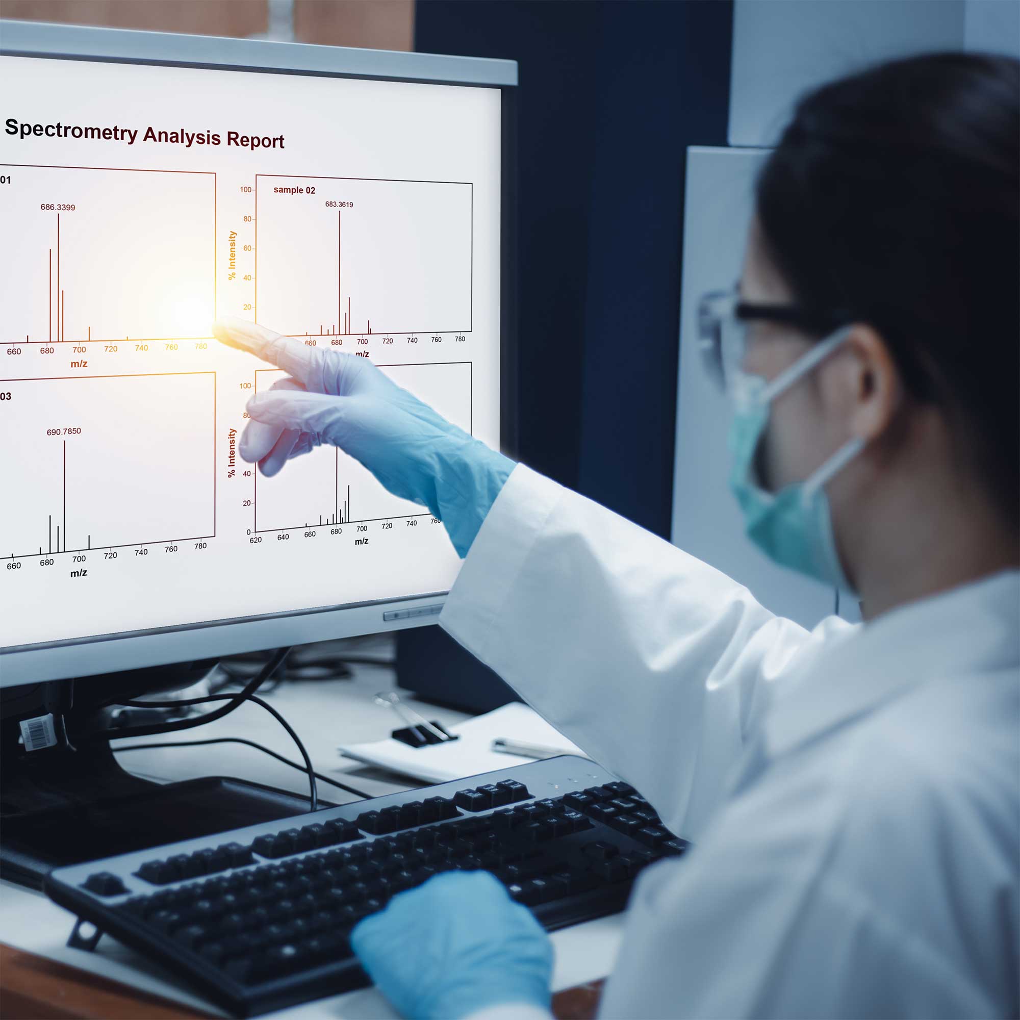
Mastering FTIR Spectroscopy: Sample Preparation and Data Analysis

Mastering FTIR Spectroscopy: Sample Preparation and Data Analysis
Fourier Transform Infrared (FTIR) spectroscopy is a powerful analytical technique used in a wide
range of fields, including chemistry, material science, pharmaceuticals, and environmental
monitoring. FTIR is essential for identifying molecular structures, detecting functional groups, and
characterizing materials by measuring the absorption of infrared radiation at different wavelengths.
To fully harness the capabilities of FTIR spectroscopy, proper sample preparation and data analysis
are crucial. In this article, we will delve into best practices for preparing samples and analyzing FTIR
data to ensure accurate and reliable results.
1. Understanding FTIR Spectroscopy
FTIR spectroscopy works by passing infrared light through a sample and measuring the absorption of
different wavelengths of light. The absorption pattern, known as the FTIR spectrum, provides
valuable information about the molecular composition and functional groups present in the sample.
The spectrum can be plotted as a graph with wavelength or frequency on the x-axis and absorbance
or transmittance on the y-axis.
FTIR is widely used for:
Identifying functional groups such as hydroxyl, carbonyl, and amine groups.
Characterizing materials like polymers, chemicals, and pharmaceutical compounds.
Analyzing contaminants and impurities in products.
Monitoring reaction progress in real-time.
For the technique to be effective, however, it’s important to prepare your sample correctly and to
analyze the data with the right methods.
2. Sample Preparation for FTIR Spectroscopy
Proper sample preparation is a critical step in obtaining reliable and reproducible FTIR results. The
preparation method depends on the physical state of the sample (solid, liquid, or gas) and its nature.
Below are common sample preparation techniques used in FTIR spectroscopy:
a. Solid Samples
For solid samples, the preparation technique used often depends on the sample's properties. Two of
the most common methods are:
KBr Pellet Method: One of the most widely used methods for solid samples is mixing the
sample with potassium bromide (KBr) and pressing it into a pellet. KBr is transparent to
infrared light, so it does not interfere with the sample's absorption spectra.
o Procedure: Weigh a small amount of the sample (typically 1-2 mg) and mix it with
approximately 100 mg of KBr powder. The mixture is then pressed under high
pressure to form a thin pellet. The pellet is placed in the FTIR instrument for
analysis.
o Considerations: Ensure that the pellet is uniform and free from cracks. Moisture can
also interfere with the spectrum, so it's crucial to keep the sample dry.
Attenuated Total Reflection (ATR): ATR is a non-destructive technique that allows you to
analyze solid samples directly without extensive preparation.
o Procedure: Place the solid sample directly on the ATR crystal, and the infrared light
is directed onto the sample surface at an angle, creating multiple internal
reflections. The spectrum is obtained from the reflected light.
o Advantages: ATR is faster, does not require complex sample preparation, and is
ideal for samples that cannot be easily dissolved or pelletized.
b. Liquid Samples
For liquid samples, FTIR spectroscopy can be performed using an optical path cell (typically a liquid
cell) with a specific path length.
Procedure: Place a few drops of the liquid sample between two infrared-transparent
windows (usually made of materials like KBr or NaCl). The sample is then placed in the
sample compartment of the FTIR instrument.
Considerations: Avoid contamination of the windows, as even small amounts of impurities
can affect the spectrum. For high absorbance liquids, you may need to use cells with shorter
path lengths.
c. Gas Samples
For gas-phase samples, a specialized gas cell with a long optical path length is used to ensure
sufficient interaction with the infrared radiation.
Procedure: The gas sample is introduced into the gas cell, and the absorption spectrum is
measured.
Considerations: Ensure that the gas is free from moisture or other interfering compounds
that may affect the spectrum.
3. FTIR Data Acquisition
Once the sample is properly prepared, the next step is data acquisition. The FTIR instrument will
scan the sample across a range of infrared wavelengths and generate a spectrum that represents the
sample's absorption at each frequency. The key to obtaining high-quality data is selecting the right
settings for the FTIR analysis.
a. Scanning Range
Most FTIR instruments are set to scan from approximately 4000 cm^-1 to 400 cm^-1. The exact
range may vary depending on the sample type. For organic compounds, scanning from 4000 cm^-1
to 600 cm^-1 is typically sufficient.
b. Resolution
The resolution of the FTIR spectrum determines how finely the frequencies are sampled. Higher
resolution allows for more detailed spectra and is particularly important when identifying subtle
functional group peaks. A resolution of 4 cm^-1 is common, but higher resolutions (e.g., 0.5 cm^-1)
are available on some instruments.
c. Number of Scans
Increasing the number of scans can improve the signal-to-noise ratio, resulting in clearer spectra.
However, it also increases analysis time. Typically, 16 to 64 scans are performed for routine analysis,
depending on the sensitivity required.
4. Data Analysis and Interpretation
Once the FTIR spectrum has been generated, the next crucial step is data analysis and interpretation.
Here’s how you can analyze your FTIR spectra effectively:
a. Identifying Key Peaks
FTIR spectra typically display characteristic peaks corresponding to the vibration frequencies of
specific functional groups in the sample. Key steps for analyzing these peaks include:
Assigning Functional Groups: The absorption peaks in the spectrum correspond to the
vibration modes of specific functional groups (such as C-H, O-H, N-H, C=O). Use a reference
table (like the Infrared and Raman Characteristic Group Frequencies) to assign functional
groups to peaks.
o For example, the O-H stretch typically appears around 3200–3550 cm^-1, while the
C=O stretch appears around 1725 cm^-1.
Peak Intensity and Shape: The intensity of a peak is related to the concentration of the
corresponding functional group, while the shape can provide insight into molecular
interactions or the presence of hydrogen bonding.
b. Subtracting Background Noise
Background noise can sometimes interfere with the clarity of the spectrum. Most FTIR software
provides tools for background subtraction, allowing you to remove noise and obtain a cleaner
spectrum. It’s also important to perform a blank scan to subtract any potential instrument-related
noise.
c. Identifying Impurities
By comparing the sample spectrum with reference spectra, you can identify impurities or
contaminants in the sample. For instance, peaks outside of the expected range or additional peaks
may indicate the presence of foreign substances.
d. Using Software for Analysis
Modern FTIR instruments come with powerful software that aids in the analysis and interpretation
of spectra. Software tools can assist in peak identification, quantitative analysis, and comparison
with spectral libraries. Some FTIR systems even offer multi-dimensional analysis, such as 2D
correlation spectroscopy, for more complex samples.
5. Common Challenges and Troubleshooting in FTIR Spectroscopy
Even with proper sample preparation and data analysis, challenges can arise during FTIR
spectroscopy. Common issues include:
a. Poor Signal-to-Noise Ratio
This can be caused by inadequate sample preparation, improper resolution settings, or
contamination. Increasing the number of scans or adjusting the gain may help improve the signal
quality.
b. Overlapping Peaks
When peaks overlap, it can be difficult to identify individual components. In such cases, advanced
analysis techniques, such as deconvolution or second derivative analysis, can help resolve
overlapping peaks.
c. Contamination
Cross-contamination from the sample handling process or the equipment can lead to distorted
spectra. Ensure that all equipment and tools are clean, and always use fresh sample holders and
windows.
Conclusion
FTIR spectroscopy is a versatile and powerful tool for molecular analysis, but to achieve accurate
results, proper sample preparation, data acquisition, and interpretation are key. By following best
practices for sample preparation, adjusting instrument settings appropriately, and using effective
data analysis methods, you can maximize the potential of FTIR spectroscopy in your research and
testing.
With Labindia Analytical’s FTIR instruments, you can ensure precise sample analysis with advanced
features, intuitive software, and reliable performance. Whether you're conducting routine material
identification or complex structural analysis, mastering FTIR spectroscopy will empower you to
uncover valuable molecular insights with confidence.
SEO Keywords:
FTIR Spectroscopy
Sample Preparation for FTIR
FTIR Data Analysis
FTIR Spectral Interpretation
FTIR Functional Group Identification
FTIR Troubleshooting
Labindia FTIR Instruments
FTIR Sample Preparation Techniques
FTIR Spectrum Analysis